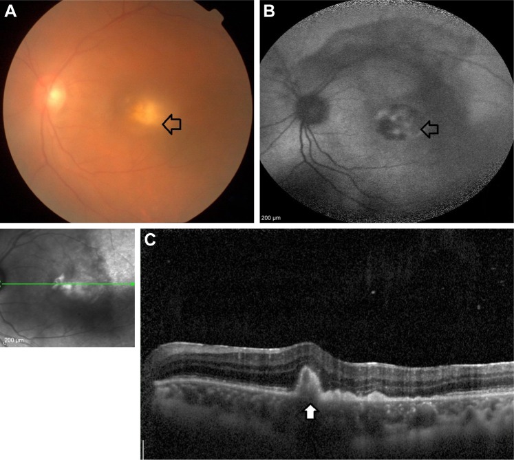
Figure 1.
Clinical and imaging features of a patient diagnosed with PVRL. (A) Left eye color fundus image of a 56-year-old male treated with oral corticosteroids for recurrent posterior uveitis with vitritis (2+). An ill-defined yellowish lesion is noted at the posterior pole of the fundus and vitreous biopsy confirmed it is a case of intraocular lymphoma (arrow with black outline). (B) Fundus autofluoroscence showing areas of hypo with hyper autofluorescence in the posterior pole of the left eye (arrow with black outline). (C) SDOCT reveals hyperreflective sub-RPE deposits characteristic of intraocular lymphoma (white arrow with black outline).
Abbreviations: PVRL, primary vitreoretinal lymphoma; RPE, retinal pigment epithelium; SDOCT, spectral domain optical coherence tomography.

