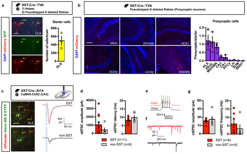
Figure 2. DLS SST-INs receive direct monosynaptic inputs from CA3.
a) SST-Cre::TVA bigenic mice were injected with helper virus (AAV8- EF1a –FLEX-HB) followed by pseudotyped G-deleted rabies virus (EnvA-SADΔG-mCherry) in the DLS. Yellow arrowheads denote starter cells, which are positive for both GFP (helper) and mCherry (rabies). Representative images for 3 independent animals. Scale bar: 50 μm. Means ± SEM; n= 3 mice per group. b) Presynaptic partners were identified in the MS/DBN, dorsal subiculum, CA1, dorsal and ventral CA3. Representative images for 3 independent animals. Scale bar: 100 μm. Means ± SEM; n= 3 mice per group. c) Example recordings of blue light-evoked monosynaptic inputs onto DLS neurons in acute slices obtained from adult SST-Cre::Ai14 bigenic mice. Clear yellow arrowheads indicate tdTomato-labeled SST INs; solid yellow arrowhead, dye fills of the recorded cell. Representative images for 4 independent animals. Scale bar: 30 μm. Traces show synaptic currents evoked in both SST-INs (top) and non-SST INs (bottom) and the 10 ms light pulse is indicated by a blue box. Individual trials are shown in gray and averages in red and black. d) Average amplitude and latency for both cell types. Means ± SEM; n= 11, 6 cells per group, unpaired Student two-tailed T-test. e) Firing properties of SST and non-SST INs tested by current injection. f) Example recordings of miniature synaptic currents in SST neurons (red) and non-SST cells (black). g) Amplitude and frequency for mEPSCs in SST and non-SST INs. Means ± SEM; n= 6, 5 cells per group, Student two-tailed T-test. All statistics detailed in Supplementary Table 1.

