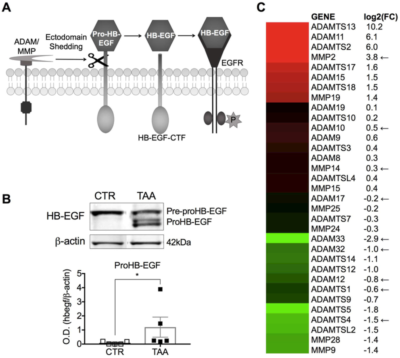
Fig. 5. Cirrhotic LSECs fail to release HB-EGF from the cytosolic membrane.
(A) Illustration of HB-EGF ectodomain shedding. (B) Western blot analysis revealed that TAA LSECs retained proHB-EGF (*P<0.05). (C) Gene expression analysis of ADAMs and MMPs indicated downregulation of 6 of the 9 sheddases involved in HB-EGF shedding in TAA LSECs (indicated by arrows). Log2(FC) represents the fold change of TAA LSECs compared to CTR LSECs. Data are mean ± s.e.m. Unprocessed original scans of blots are shown in Supporting Fig. S6. See Supporting Table S3 for numbers of replicates and details of statistical analysis.

