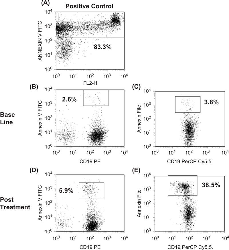
Figure 2.

Detection of apoptosis by annexin V staining in B cells. (A) Annexin V staining (y-axis) in positive control. (B) Low level (2.6%) apoptotic B cells (in box) staining positive for CD19 (x-axis) and annexin V (y-axis). (C) Low level (3.8%) apoptotic B cells (in box) staining positive for CD19 (x-axis) and annexin V (y-axis). (D) Moderate level (5.9%) apoptotic B cells (in box) staining positive for CD19 (I-axis) and annexin V (y-axis). (E) High level (38.9%) apoptotic B cells (in box) staining positive for CD19 (x-axis) and annexin V (y-axis). Patient 10 is shown in (B) and (D) while patient 17 is shown in (C) and (E).
