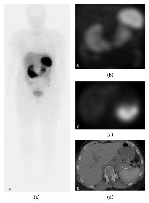
Figure 2.

24-hour 111In-pentetreotide scan. Anterior whole body planar image (a), MIP reconstruction of SPECT acquisition (b), axial SPECT image (c), and fused axial SPECT/CT image (d) show normal biodistribution of the radiotracer and no uptake in the pancreatic tail mass, ruling out a splenule.
