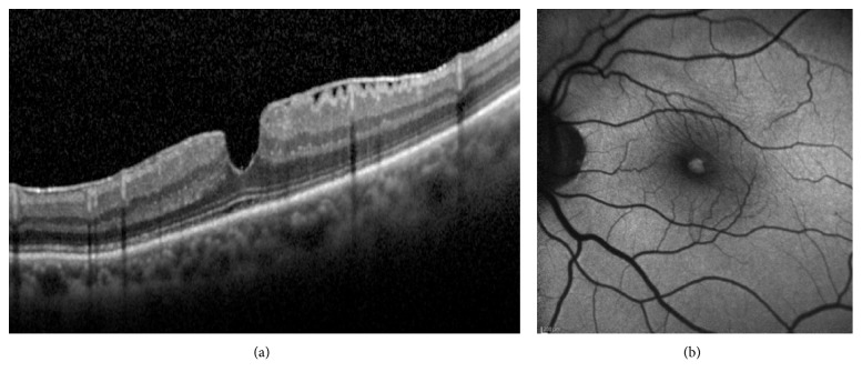
Figure 4.
(a) SD-OCT demonstrating an epiretinal membrane, irregular foveal contour with steep edges, no break in the inner fovea or dehiscence of the inner retina from the outer retina. According to OCT criteria, a diagnosis of MPH should be established. Enhanced visualization of Henle's fiber layer fails to determine with the certainty if there is a loss of tissue in the center of the foveola. (b) Corresponding fundus AF image showing a well-defined lack of central macular pigment. A diagnosis of LMH should be made.

