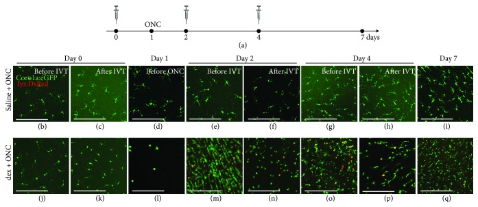
Figure 3.
Local immunosuppressive treatment with dex induces an exaggerated inflammatory response to subsequent optic nerve injury. (a) Schematic representation of the experimental setup. Intravitreal injection (IVT) of dex (or saline) is performed three times, at days 0, 2, and 4. ONC is performed at day 1. Retinas are harvested just before and at 6 hours after each injection (days 0, 2, and 4), just before the moment of ONC (day 1), and at day 7, to assess their number and morphology in the inner retina. (b-i) Intravitreal injection of saline has no effect on the retinal microglia appearance before or after ONC. However, from day 4 onwards (i.e., 3 dpi), a restricted increase in the number of microglia/macrophages can be observed, as we have shown before [21]. (j-q) As a result of the first intravitreal dex injection at day 0, the inner retina is almost completely devoid of microglia at the moment of ONC (day 1). Nevertheless, at day 2 (before the second injection), the number of microglia/macrophages increases drastically, compared to the previous time points, and to the saline + ONC fish. Neutrophil infiltration is observed as well (yellow). This increase is partly counteracted by the second dex injection. Indeed, after the injection at day 2, the number of microglia/macrophages and neutrophils is reduced. At day 4, the number of innate immune cells increased again, and subsequent dex injection temporarily decreased this response once more. At day 7, the number of microglia/macrophages is again higher than right after the injection at day 4 in the dex + ONC group. Additionally, there are still more innate immune cells in the dex + ONC than in the saline + ONC group at 7 days post-ONC. Scale bars: 50 μm.

