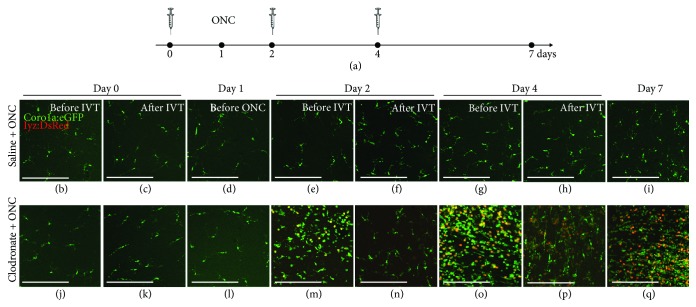
Figure 5.
Local immunosuppressive treatment with clodronate liposomes induces an exaggerated inflammatory response to subsequent optic nerve injury. (a) Schematic representation of the experimental setup. Intravitreal injection of clodronate liposomes (or saline) is performed three times, at days 0, 2, and 4, and ONC is performed at day 1. The inflammatory response is assessed just before and 6 h after each injection (days 0, 2, and 4), just before the moment of ONC (day 1), and at day 7. (b-i) Intravitreal injection of saline does not affect retinal microglial appearance before nor after ONC. A restricted increase in the number of microglia/macrophages can be observed from day 4 onwards (i.e., 3 dpi), as we have shown before [21]. (j-q) Although full microglial depletion is not yet obtained at day 1, ONC induces a prominent increase in the number of retinal microglia/macrophages at day 2 (before the second injection) in the clodronate + ONC retinas, as compared to saline + ONC retinas. Neutrophil infiltration is apparent as well in the clodronate + ONC group at this point. This inflammatory reaction is partially counteracted by subsequent application of clodronate liposomes at day 2. However, this suppression is clearly temporary as the number of innate immune cells is again massively increased before the third clodronate injection at day 4. Again, this striking inflammatory response can be blocked by a third clodronate injection. Still, at day 7 the number of microglia/macrophages is much higher than seen in the saline + ONC group at the same time point. Scale bars: 50 μm.

