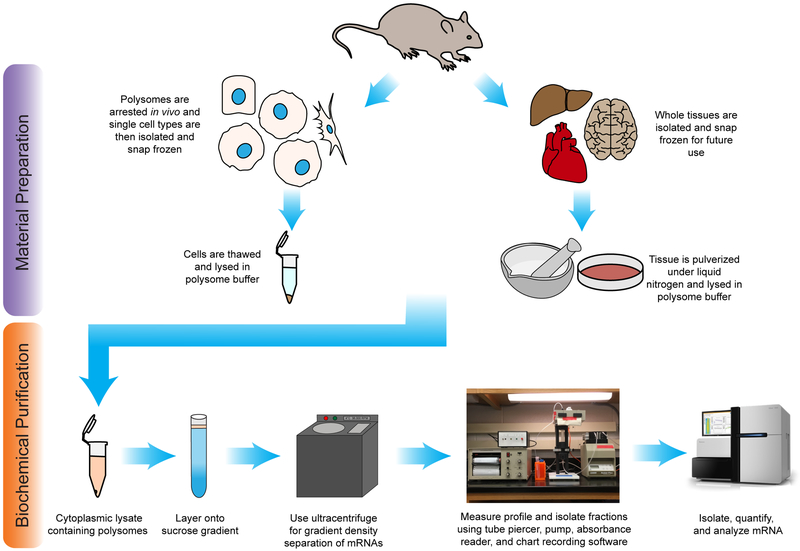
Figure 1. Experimental schematic.
The experimental workflow of polysome profiling from mammalian tissues or isolated cell-types following by next-generation sequencing analysis. Either whole tissue or single cell-types are isolated from the animal. The cells are lysed, nuclei and cell debris are removed, and the cytoplasm is layered onto a sucrose gradient. Following centrifugation, the gradient is flowed through an absorbance detector and the profile is collected. mRNA can then be isolated from fractions for downstream analysis.

