Figure 6.
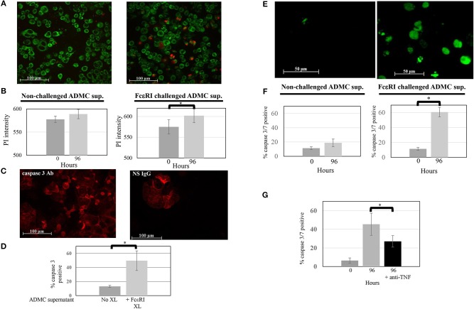
Mediators from FcεRI-challenged ADMC induce SK-BR-3 cell killing. (A) ADMC (1.3 × 10⁶) were challenged with optimal concentrations of anti-FcεRI stimuli (70% release) for 24 h and supernatants (XL media; no cells) from these ADMC were incubated with the MitoTracker™ green-stained SK-BR-3 (105) in culture medium containing optimal concentrations of PI and images taken before (left) and after (right) 96 h. (B) Quantification of overall PI fluorescence before and after incubation. The increased number of red cells indicates breast cancer cell death as indicated by the PI (red) and quantified in showing overall PI fluorescence before and after incubation. Graph represents average PI intensity from two separate experiments (±SD; *p = 0.0008). (C) Mediators from FcεRI-challenged ADMC induce human breast cancer cell apoptosis. The same media from anti-FcεRI challenged ADMC were incubated with SK-BR-3 (105) for 72 h, cytospins prepared, fixed, and incubated with Alexa Fluor 647 labeled, anti-human caspase 3 (left) or Alexa Fluor 647 labeled, isotype control Ab for caspase 3 (right). Representative panels are shown. (D) Quantification of overall Alexa FluorTM 647 fluorescence before and after incubation with supernatants from FcεRI activated ADMC. *p = 0.01 SK-BR-3 apoptosis comparing anti-HER2/neu IgE-sensitized cells at day 0 vs. day 4 from three experiments. (E) Mediators from FcεRI-challenged ADMC induce SK-BR-3 cell killing measured by caspase 3/7. ADMC (1.8 × 10⁶) were challenged with optimal concentrations of anti-FcεRI stimuli (63% release) for 24 h and supernatants (no cells) from these ADMC were incubated with SK-BR-3 (105) and caspase 3/7 green images taken before (left) and after (right) 96 h. The increased number of green cells indicates breast cancer cell death as indicated by the caspase 3/7 and quantified by counting live vs. dead cells before and after incubation. (F) Graph represents average percentage of cells from two separate experiments (±SD; *p = 0.002). (G) Blocking TNF-α significantly reduces SK-BR-3 cell death. SK-BR-3 were treated and quantified as in (C) except anti-TNF-α Ab were added during the incubation time. *p = 0.028 Decrease in SK-BR-3 cell death when anti-TNF-α Ab are added to the supernatants from anti-FcεRI stimulated ADMC.

