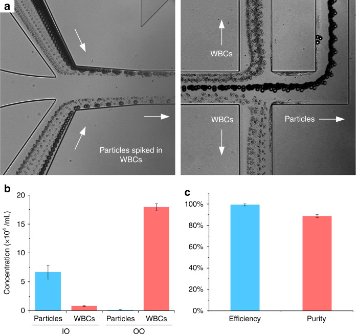
Fig. 5. Separation of spiked particles from white blood cells (WBCs).
a Bright field images of particles and white blood cells at input and output. Particles were sized 18.7 μm in diameter which is larger than most of WBCs and exiting from the central output channel. b Concentrations (N = 3) of WBCs and particles collected from inner outlet (IO) and outer outlet (OO). c Efficiency and purity for particles collected from IO. Total flow rate was 300 μL/min and flow rate ratio was 1:2 between sample and buffer

