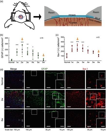
Fig. 1.
Soft cranial window installation and the time course of reactive astrocyte and microglial expression postsoft cranial window installation. (a) Schematic of the soft cranial window installation with PDMS. (b) and (c) Quantification of GFAP(+) cells (reactive astrocyte) and Iba-1(+) cells of the cortex within the soft cranial window area at different postsurgery time points ( in each group). (d) Immunohistochemistry of normal, 2 weeks postsurgery, and 6 weeks postsurgery mice. GFAP levels of astrocytes and microglia were highly activated in the brain of the animals at 2 weeks postsoft cranial window installation (*, **, ***).

