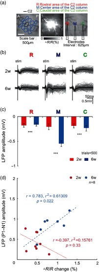
Fig. 5.
LFP at 2 and 6 weeks postsoft cranial window installation. (a) In vivo LFP recording set up with C2 whisker single deflection. Three electrodes recorded LFPs simultaneously in the center of the C2 column and in both the rostral and caudal directions. (b) LFP following C2 whisker single deflection (trials = 100). (c) Peak amplitude of LFP following C2 whisker single deflection (trials = 500, all ). (d) Correlation of LFP amplitude in center of column and the peak value of CBV change.

