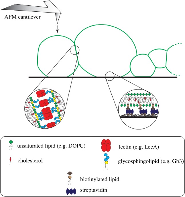
Figure 5.

Schematic illustration of prototissues and possible application of micromanipulation techniques on prototissues. The schematic illustration depicts protocell–protocell junctions and protocell–substrate adhesion in a prototissue (not to scale). The protocell–protocell adhesion is regulated by lectin–glycan interactions, while the protocell–substrate is controlled by biotin–streptavidin attachment. The atomic force microscopy technique might be applied to prototissues to measure membrane physical parameters as discussed in §2 for single vesicles. (Online version in colour.)
