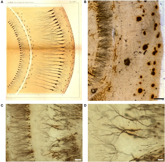
Figure 6.
Drawing (A) and images (B–D) from a Golgi-impregnated “vertical section” through the pes Hippocampi major (Ammon's horn) of the rabbit. (A) The drawing is Plate XXI from Golgi (1885), and, as for Figure 5, the translation is provided by Bentivoglio and Swanson in Golgi et al. (2001). In the figure legend Golgi described a “ventricular epithelium,” composed by cells “strikingly analogous to. neuroglial cells,” a “convoluted gray layer,” and “small nerve cells of the fascia dentata.” (B–D) Images from a Golgi's slide. Scale bars: 200 μm in (B), 30 μm in (C), 50 μm in (D).

