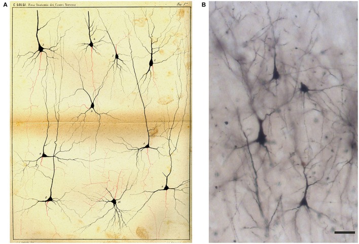
Figure 7.
Drawing (A) and images (B) of Golgi-impregnated neurons of the cerebral cortex. (A) The drawing is Plate I from Golgi (1885). In the legend Golgi stated that the plate, illustrating “some types of ganglion cells in the cerebral cortex,” is especially destined to show the origin and branching of the axon (“the only nervous prolongation of each ganglion cell”); the cells are from the frontal (“anterior central”; cells 1, 2, 4, 5, 9, 10) and occipital (cells 3, 6–8) human cerebral cortex. The legend states that, on the basis of their axonal ramifications, cells 1 and 3 illustrate examples of the first type of neurons (currently named as “Golgi type I,” see text) and cell 2 an example of the second type (currently “Golgi type II”). The legend also states that the axon could not be followed because it became too thin “destined to get lost in the diffuse net.” Scale bar in (B): 30 μm.

