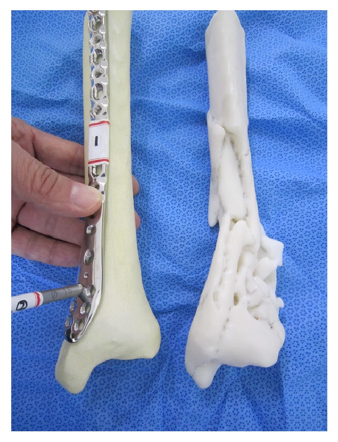
Figure 4.

Surgeons were given time to use these 3D models to study the fracture configuration and to simulate the placement of the plates on the fractured tibia considering screw trajectories in the plate for fixation of fracture fragments. The normal-side tibia model was provided to simulate the fractured tibia after reduction. Surgeons were then asked again to select the plates most suitable for the two distal tibia fractures.
