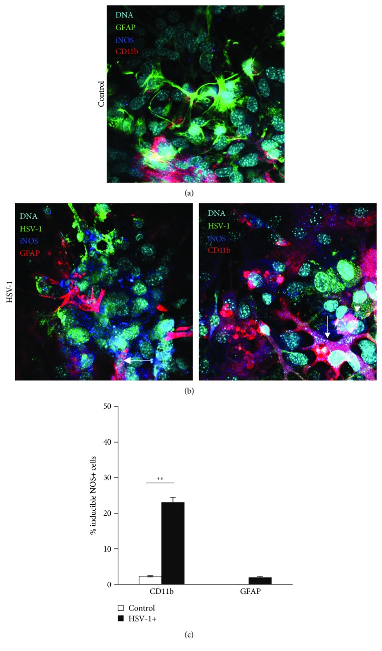
Figure 2.
Cells positive for iNOS in HSV-1-infected mixed cultures. Representative confocal images of mixed glial cultures uninfected (a) or HSV-1 infected (b) at 24 h p.i. and stained for HSV-1, astrocytes (GFAP+), microglia (CD11b+), and iNOS (inducible NOS). Nuclei were counterstained with DAPI. (c) Percentage of CD11b+ and GFAP+ cells positive for iNOS accessed by flow cytometry in HSV-1-infected and control uninfected mixed glial cultures. Means are expressed as mean ± SEM for n = 3; ∗significant differences with p ≤ 0.05, while ∗∗p ≤ 0.01.

