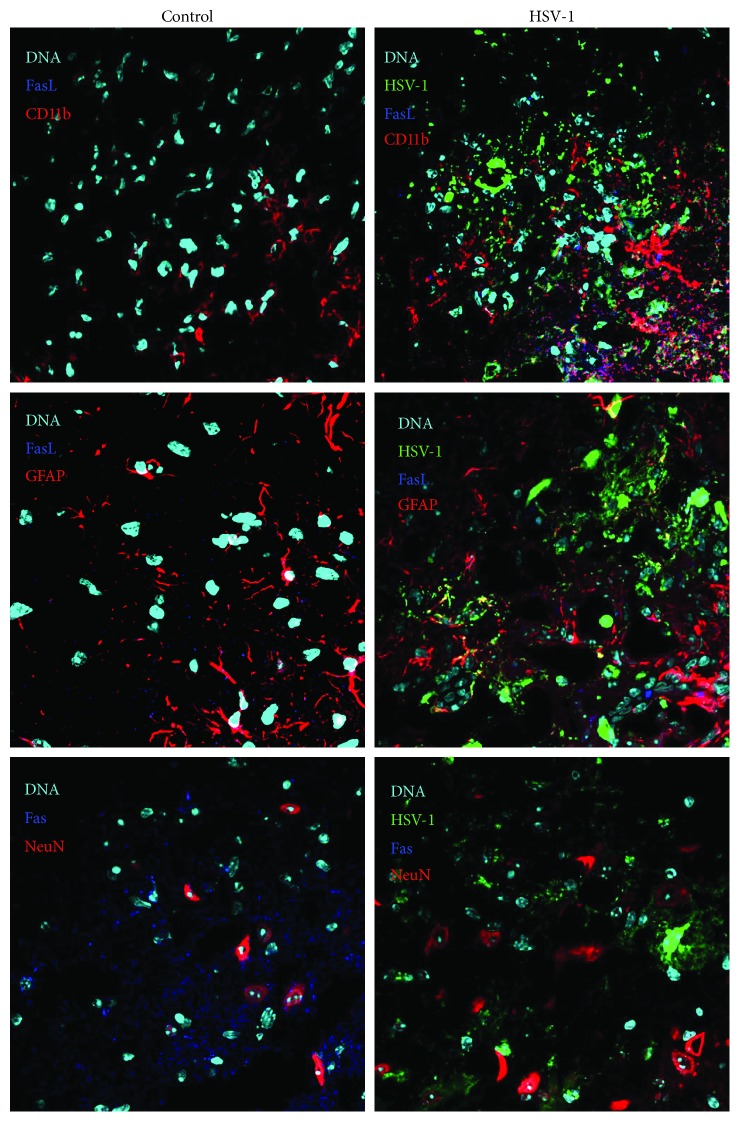
Figure 6.
Microglia are the main source of FasL in HSV-1-infected brains. Representative confocal images of the brains obtained at day 5 p.i. with HSV-1. Tissue sections from brain stems were stained for microglia (CD11b+), astrocytes (GFAP+), neurons (NeuN), Fas, and FasL. Nuclei were counterstained with DAPI.

