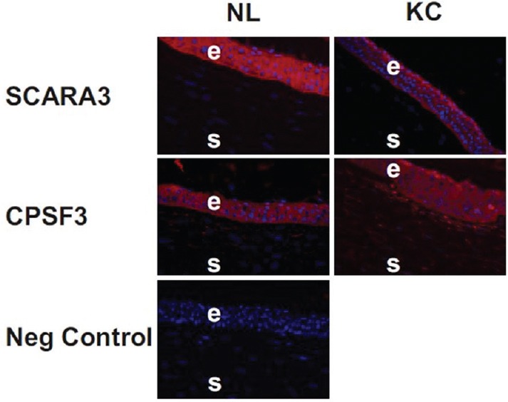
Figure 4.

Immunohistochemistry shows SCARA3 staining in NL and KC corneas. The epithelial cells have a distribution of SCARA3 throughout the cytoplasm of the NL and KC corneas. The CPSF3 protein staining is found within the cytoplasm of the epithelial cells and stromal cells. No staining was observed when only the secondary antibody (IgG) was used on the tissue sections (negative control). NL, normal; KC, keratoconus, E, epithelial; S, stroma.
