
Figure 3.
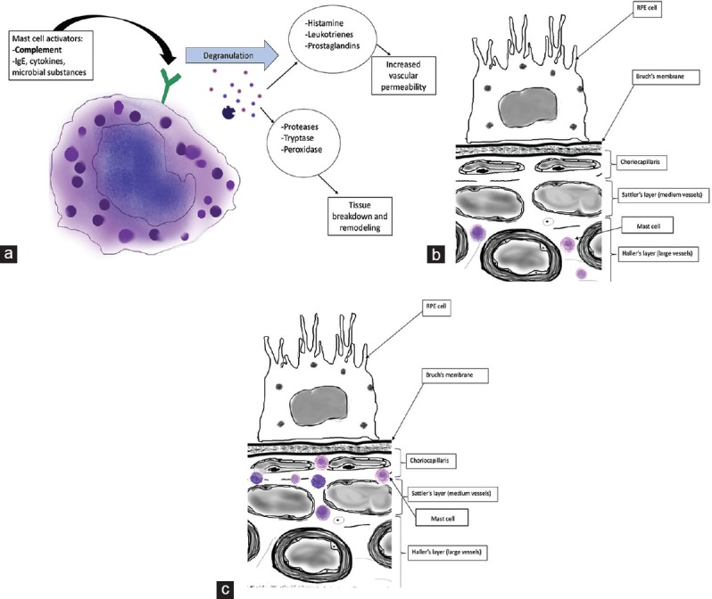
(a) Schematic of mast cell activation and degranulation. MCs are activated by complement, cytokines, etc., Granule contents include proteases that cause tissue breakdown and remodeling and vasoactive inflammatory mediators such as histamine and prostaglandins. MC activation in the choriocapillaris may contribute to CNV.[32] (b) Mast cell location in choroid of normal eye. MCs remain in Sattler's and Haller's layers.[30] (c) Mast cell location in the choroid of an eye with AMD. MCs migrate to the choriocapillaris.[32]