
Figure 4.
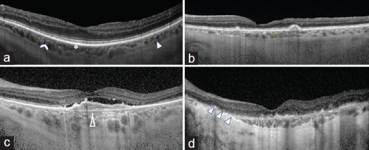
Representative SD-OCT imaging of the choroid in AMD progression. (a) normal control with all three choroidal layers visible: choriocapillaris (asterisk), Sattler's layer (chevron) and Haller's layer (solid arrow head). (b) Intermediate AMD with drusen and thinning of the choroid compared with normal control. (c) Late AMD with CNV and atrophy of the choriocapillaris (hollow arrow head). (d) Late AMD with GA and severe atrophy of the choroid (triple arrow heads).