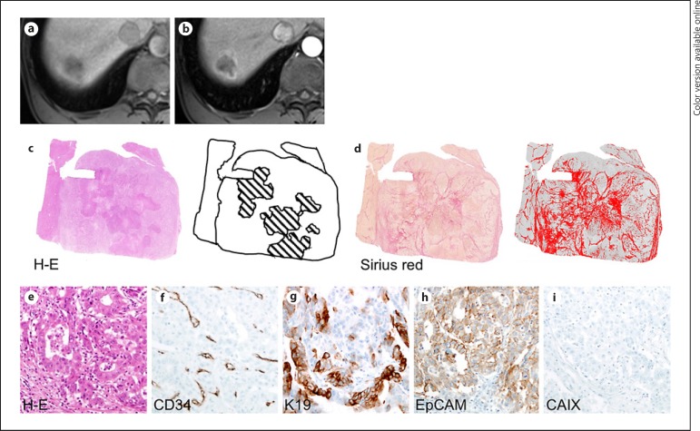
Fig. 3.
A 73-year-old male with hepatitis B virus-related cirrhosis and hepatocellular carcinoma with irregular rim-like enhancement (IRE-HCC). A 3-cm tumor in segment 8 of the liver shows low-signal intensity on precontrast T1-weighted imaging (a) and IRE in the arterial phase (b) of gadoxetate-enhanced MRI. There is necrosis in its central portion (c) (hematoxylin-eosin [H-E] stain, necrosis is hatched in black), and a high proportion of fibrosis is present (d) (Sirius red stain; fibrous stroma is colored in red). On histopathologic analysis, HCC demonstrated abundant fibrous stroma-intervening cords of tumor cells (e, H-E stain), mixed sinusoid-like and capillary-like microvascular pattern with low microvascular density (199/mm2) (f), positive expression of K19 (g) and EpCAM (h), and negative expression of carbonic anhydrase IX (CAIX) (i). Original magnification, ×200.

