Figure 1.

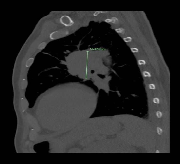
Sagittal computed tomography scan of the chest demonstrating a 5.6 cm left superior hilar mass suspicious for pulmonary malignancy.

Sagittal computed tomography scan of the chest demonstrating a 5.6 cm left superior hilar mass suspicious for pulmonary malignancy.