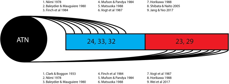
FIGURE 3.
Thalamocingulate tract termination in cingulum. Diagrammatic overview of the quantity of studies showing regions of thalamic termination once enclosed within the cingulum bundle. Brodmann area 24, 33, and 32 (shown in blue) correspond to the anterior cingulate cortex. Brodmann area 23 and 29 (shown in red) correspond to the posterior cingulate cortex and retrosplenial cortex. ATN, anterior thalamic nucleus.

