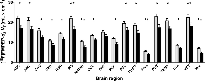
Figure 1.

Distribution volume (VT) of [18F]FMPEP-d2 (a measure of cannabinoid CB1 receptor density) is lower in male tobacco smokers (black bars, n = 18) than in nonsmokers (gray bars, n = 28) in both cortical and subcortical regions. Values are estimated marginal means from the repeated-measures analysis of variance and are adjusted to an average body mass index of 26.8 kg/m2. Error bars are standard error of the mean. *p < .05; **p < .005; post hoc contrasts of marginal means from analysis of variance. ACC, anterior cingulate cortex; AMY, amygdala; CAU, caudate nucleus; CER, cerebellum; HIPP, hippocampus; INS, insula; MIDBR, midbrain; OCC, occipital cortex; PAR, parietal cortex; PCC, posterior cingulate cortex; PFC, prefrontal cortex; PHIPP, parahippocampal gyrus; PUT, putamen; TEMP, lateral temporal cortex; THA, thalamus; VST, ventral striatum; WM, white matter.
