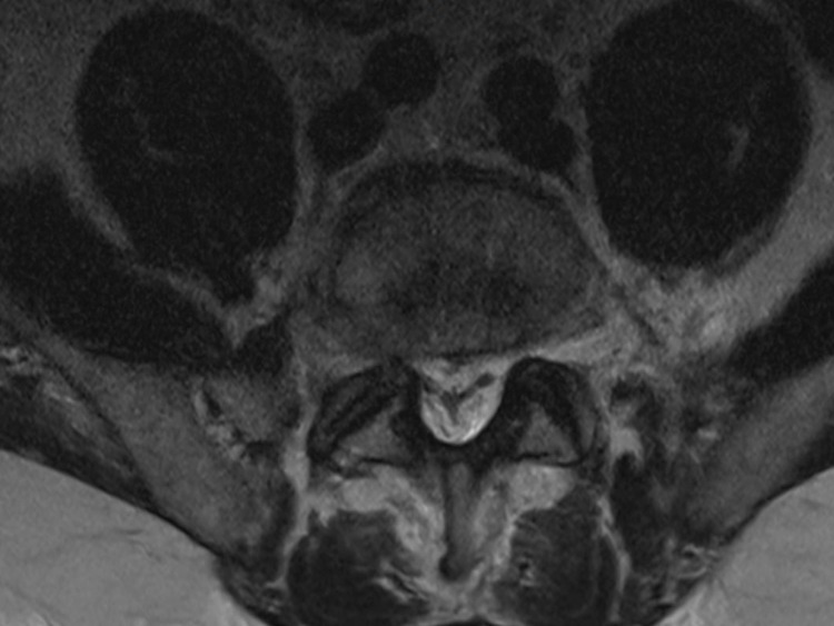
Figure 3.
Axial T2-weighted MRI scan of the lumbar spine post nerve root decompression at level L5/S1. Image demonstrates the characteristic ‘Y’ sign configuration of thecal sac compression due to compression by epidural fat, as first described by Kuhn et al, indicating stage three spinal epidural lipomatosis.10

