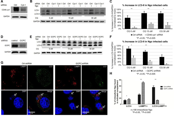
Fig 3. Autophagic flux in Ngo infected cells is mediated by CD46-cyt1.
(A)Representative immunoblot showing CD46-cyt1 and GAPDH in ME180 cells treated with control (Ctrl) or CD46-cyt1 (Cyt-1) siRNA. GAPDH in each sample was used as the internal control. (B)Representative immunoblot showing LC3-I, LC3-II and GAPDH in ME180 cells treated with Ctrl or CD46-cyt1 (Cyt-1) siRNA, followed by treatment with 0, 15 or 30 μM CQ, and mock-infected or infected with Ngo at an MOI of 10 for 4 h. (C)Densitometry quantification of immunoblots from 4 independent experiments as described in (B). For each condition, LC3-II/GAPDH levels in infected cells were normalized to LC3-II/GAPDH levels in mock-infected controls. (D)Representative immunoblot showing GOPC and GAPDH in cells transduced with lentivirus containing either control shRNA or GOPC shRNA. (E)Representative immunoblot showing LC3-I, LC3-II and GAPDH in cells transduced with lentivirus containing control (Ctrl) or GOPC shRNA and mock-infected or infected with Ngo for 4 h at an MOI of 10. (F)Densitometry quantification of immunoblots from 5 independent experiments as described in (E). For each condition, LC3-II/GAPDH levels in infected cells were normalized to LC3-II/GAPDH levels in mock infected controls. (G)Representative SIM microscopy image of an ME180 cell treated with Ctrl or GOPC shRNA, infected with Ngo for 4 h at an MOI of 10, and stained for LAMP1 (red), LC3 (green), and DAPI (blue). Arrowheads show intracellular Ngo in intracellular compartments. (H)Quantification of intracellular Ngo found in LC3+, LAMP1+ or LC3+LAMP1+ compartments 4 hpi, in cells treated with Ctrl or GOPC shRNA.

