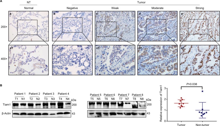
Figure 1.
Tiam1 protein expression in lung adenocarcinoma tissues.
Notes: (A) IHC staining for Tiam1 protein expression in lung adenocarcinoma. (a) Negative Tiam1 staining in normal lung tissues. (b) Negative Tiam1 staining in normal lung adenocarcinoma tissues. (c) Weak Tiam1 staining in lung adenocarcinoma. Tiam1 protein was mainly detected in the cytoplasm. (d) Diffuse and moderate Tiam1 staining in lung adenocarcinoma tissues. Protein signals were detected in both cytoplasm and cell nuclei. (e) Diffuse and strongly positive Tiam1 staining in lung adenocarcinoma tissues. Protein signals were revealed mainly in the nuclei. (a1–e1) Indicate higher magnification of the selected area in (a) to (e), respectively (original magnification, a–e: 200×; a1–e1: 400×). (B) Western blot analysis of Tiam1 expression value in tumor vs non-tumor tissue, and Tiam1 was significantly overexpressed in tumor tissue (P=0.0378).
Abbreviations: IHC, immunohistochemistry; NT, non-tumor; T, tumor; N, non-tumor.

