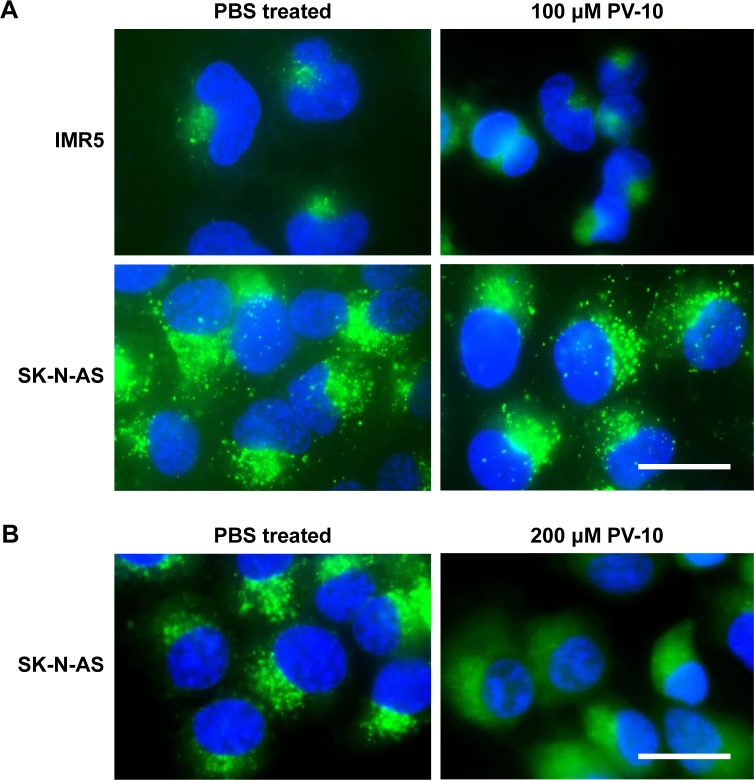
Figure 3.
PV-10 disrupts lysosomes.
Notes: Live cells were stained with the nucleic acid stain Hoechst 33342 and LysoTracker green DND-26, which concentrates and fluoresces in acidic organelles, and observed by fluorescence microscopy. (A) Neuroblastoma cell lines IMR5 and SK-N-AS were treated with either PBS (vehicle control) or 100 µM PV-10 for 6 hours. (B) Neuroblastoma cell line SK-N-AS was treated with either PBS (vehicle control) or 200 µM PV-10 for 6 hours. Scale bars = 20 µm. Data presented are representative of three separate experiments.

