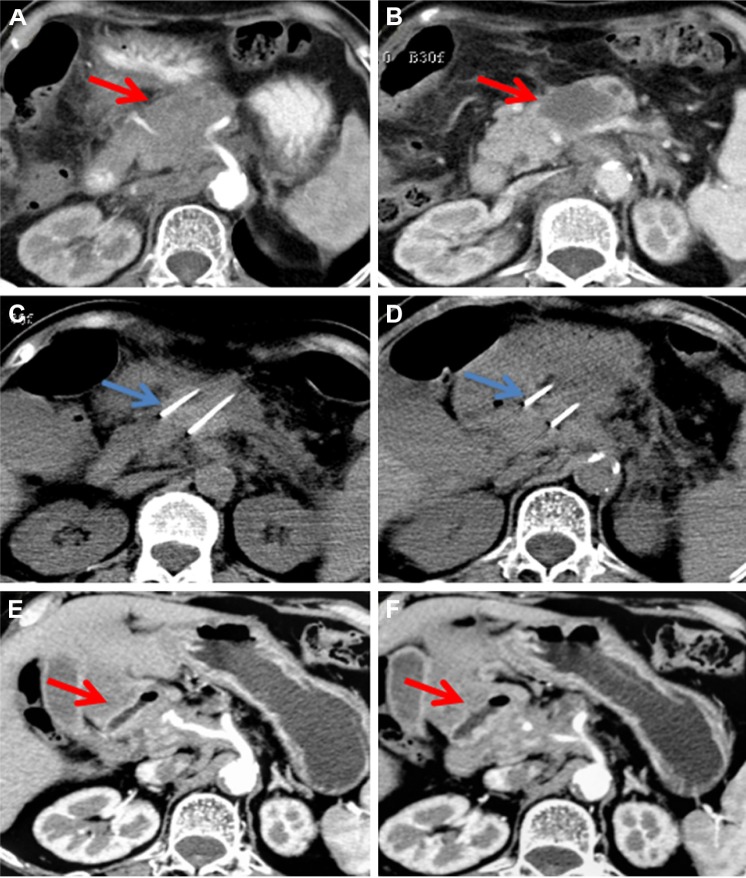
Figure 2.
A 70-year-old woman with T4N1M0, stage III, pancreatic head and neck carcinoma.
Notes: Enhanced computed tomography shows a pancreatic tumor measuring 6.9×4.8 cm (A, B). Irreversible electroporation (IRE) is performed (C, D). At 3 months after IRE, the tumor has shrunk to 3.5×2.1 cm, and vascular retention is noted (E, F). The red arrows indicate the tumor and the blue arrows indicate the IRE probes.

