Figure 1.
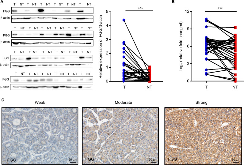
FGG is significantly upregulated in HCC tumor tissues.
Notes: (A) Western blot analysis of FGG expression in 24 pairs of HCC tumor tissues and their adjacent non-tumorous tissues; P=0.0005, paired t-test. (B) RT-qPCR analysis of FGG mRNA expression in another sample set containing 35 pairs of HCC tumor samples and their non-tumorous tissues; P=0.0018, paired t-test. (C) Different FGG expression levels in HCC tumor tissues. FGG expression was categorized into three groups: weak – weak positive staining (1+); moderate – moderate positive staining (2+); and strong – strong positive staining (3+). ***P<0.001.
Abbreviations: FGG, fibrinogen gamma chain; HCC, hepatocellular carcinoma; NT, non-tumor sample; T, tumor sample; RT-qPCR, reverse transcription quantitative PCR.

