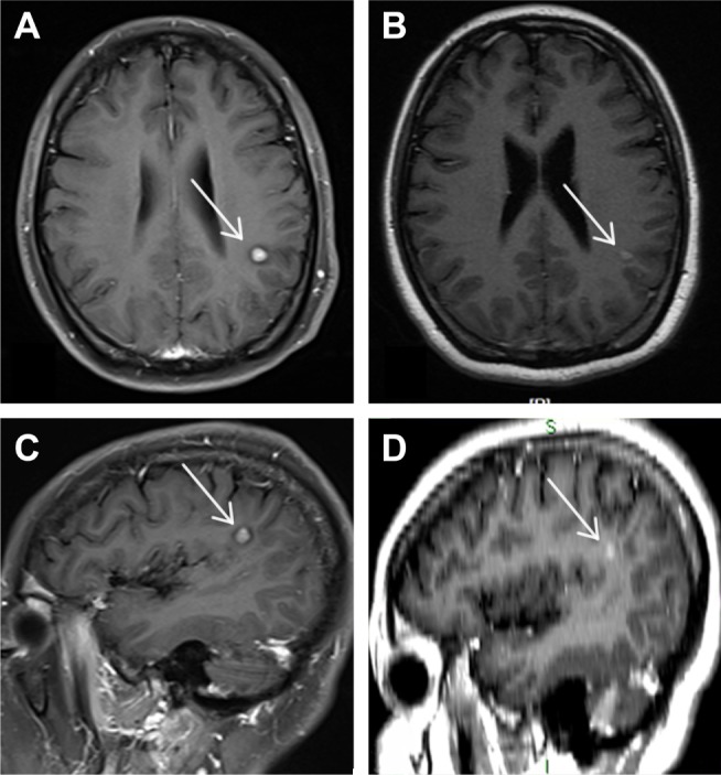
Figure 2.

Enhanced MRI of a 6 mm slice at the start of the treatment with fulvestrant showing a single metastatic lesion on the left parietal lobe (A, C) vs a repeat MRI of the same patient showing a gross reduction in the size of the metastatic lesion of the brain 1 month after administering the first dose of fulvestrant (B, D).
Notes: (B) is from a positioning MRI of a 3 mm slice, and (D) is from three- dimensional reconstruction MRI images of a 3 mm slice. The arrows in images A and C indicated the metastatic tumors.
Abbreviation: MRI, magnetic resonance imaging.
