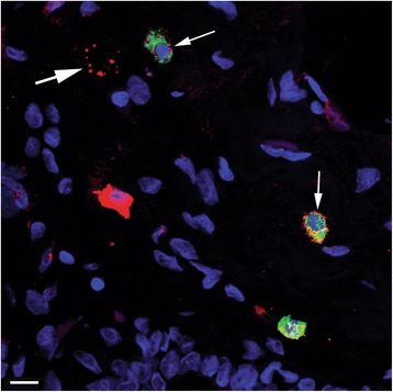
Fig. 4.

A confocal image of double immunofluorescence labeling of tryptase and SSEA3. The image is a “maximum intensity” digital composite from 12 focal planes showing merged channels from dIF labeling of tryptase (green) and SSEA3 (red), together with DAPI nuclear staining (blue). Double-labeled tryptase+ SSEA3+ round/oval cells (thin arrows) and a SSEA3+ degranulated cell (thick arrow) are indicated. Scale bar 10 μm
