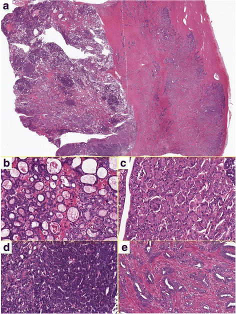
Fig. 2.

Hematoxylin and eosin staining. a Panoramic view of the tumor. The left half of the tissue is the intracystic component, and the right half is the invasive component in the myometrial layer. b Relatively uniform tubules containing dense eosinophilic secretion is characteristic. c Tumor cells within the eosinophilic cytoplasm are lined in an acinar fashion. d Tumor cells show a very high nuclear-cytoplasmic ratio in a solid pattern. e Invasive carcinoma in the myometrium. Much higher-grade carcinoma similar to conventional endometrioid adenocarcinoma invades the myometrial layer
