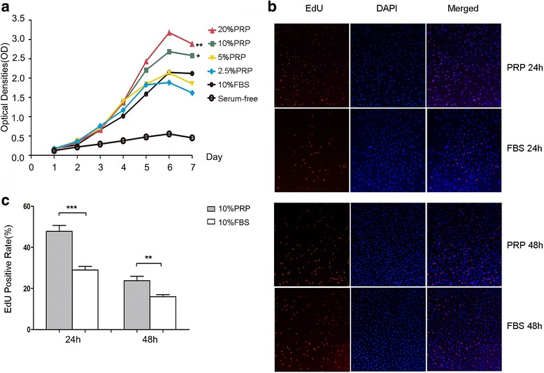
Fig. 1.

PRP promoted MenSCs proliferation. a CCK8 assay detected proliferation of P4 MenSCs cultured with different concentrations of activated PRP or 10% FBS (n = 3). b Immunofluorescence analysis of EdU+ MenSCs under stimulation of 10% PRP and 10% FBS for 24 h and 48 h (red: DNA replicating, blue: nuclei, n = 6). c Statistical analysis of EdU+ cell rate of each group. MenSCs cultured with 10% PRP showed higher proliferation rate for both 24 h and 48 h (P < 0.01). CCK8 was analyzed by one-way ANOVA test. Data was mean ± SEM, *P < 0.05, **P < 0.01, ***P < 0.001 for two-tailed t test
