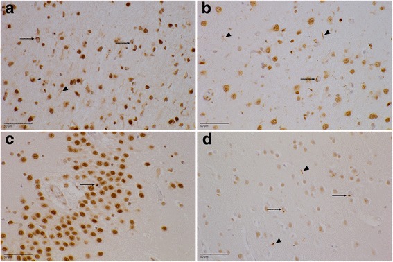
Fig. 4.

Transactive response DNA-binding protein (TDP) pathology (paraffin-embedded sections stained with antihyperphosphorylated TDP-43 antibody). a DR25.5 area 6. There is a moderate amount of neuronal intracytoplasmic inclusions (NCIs) (arrows), mainly in the second cortical layer. The dystrophic neurites (arrowhead) are more evenly spread throughout the entire cortex. b DR2.3 superior temporal gyrus. Mild to moderate TDP-43 proteinopathy type A with NCIs (arrow) and dystrophic neurites (arrowheads) can be seen. c DR28.1 dentate gyrus. There are scarce NCIs (arrow) in the granular layer. d DR31.1 area striata. A mild amount of NCIs (arrows) and dystrophic neurites (arrowheads) in the second cortical layer can be seen
