Fig. 3.

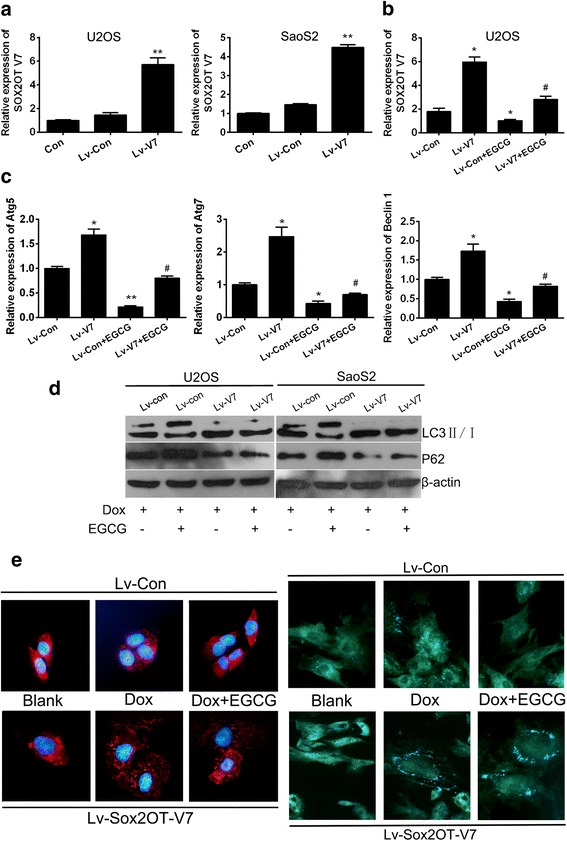
EGCG inhibited DOX-induced autophagy by targeting SOX2OT V7 partially. SOX2OT V7 gain-of-function cell models were constructed by lentivirus infection. a The relative mRNA levels of SOX2OT V7 in OS cells were checked by qRT-PCR. ** p < 0.01 vs. Lv-Con. b qRT-PCR was performed to detect the SOX2OT V7 in U2OS cells treated with or without EGCG. Results shown are representative of three independent experiments and error bars indicate SE. * p < 0.05 vs. Lv-con, # p < 0.05 vs. Lv-con treated with EGCG. c qRT-PCR was performed to detect the mRNA level of autophagy associated genes in V7 over-expressed U2OS cells. Results shown are representative of three independent experiments and error bars indicate SE. * p < 0.05 vs. Lv-con, # p < 0.05 vs. Lv-con treated with EGCG. d Western blots were performed to detect the expression of LC3 and P62 in V7 over-expressed U2OS and SaoS2 cells with different treatment. e LC3 immunofluorescence (the left half) and MDC staining (theright half), LC3 puncta distribution and MDC signals were visualized by confocal fluorescence microscopy, and representative images are shown. The images were captured at 400 x magnification
