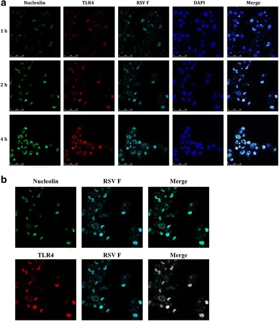
Fig. 5.

Co-localization of the RSV F protein with the cell surface receptors TLR4 and nucleolin and internalization of the RSV F protein into N2a cells were examined by laser confocal microscopy. N2a cells were fixed and stained with fluorescence-labeled antibodies against TLR4 (red), nucleolin (green) and the RSV F protein (cyan) at different time points after RSV infection. Simultaneously, N2a cell nuclei were counterstained with DAPI. a The RSV F protein co-localized with both TLR4 and nucleolin at 1 h pi, 2 h pi and 4 h pi; b The RSV F protein co-localized with TLR4 and nucleolin in N2a cells at 2 h pi; nucleolin is labeled with Alexa 488 (green fluorescence), TLR4 is labeled with Alexa 555 (red fluorescence), and F protein is labeled with Alexa 647 (cyan fluorescence). Co-localization was evaluated using an analytic tool from the LAS AF software package
