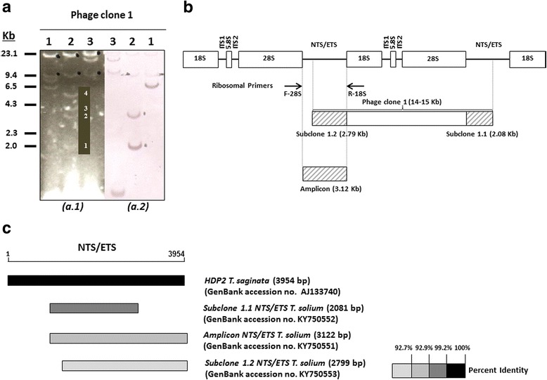
Fig. 2.

DNA sequence organization and similarities of the T. solium HDP2 fragment. a a.1: Restriction fragment patterns for T. solium recombinant phage clone 1 DNA digested by SacI (Lane 1); SalI (Lane 2); XbaI (Lane 3); a.2: Southern blot: T. saginata HDP2 DNA sequence digoxigenin-11-dUTP-labeled hybridization with T. solium recombinant phage clone 1 DNA digested by SacI (Lane 1); SalI (Lane 2); XbaI (Lane 3). b Diagram showing the genomic organization of the T. solium HDP2 subclones 1.1 and 1.2 and the T. solium HDP2 amplicon, with respect to the structure of the ribosomal DNA repeats (-18S-ITS1-5.8S-ITS2-28S-NTS/ETS-). c Physical alignment of Taenia HDP2 (NTS/ETS) fragments. The NTS/ETS ribosomal repeat is represented by a line, with the T. saginata HDP2 unit as the reference sequence. The boxes in grayscale correspond to the percent homology with the T. saginata HDP2 fragment (black box represents 100% identity). T. solium subclones 1.1 and 1.2 and the ribosomal amplicon are represented by boxes in different shades of gray according to similarities
