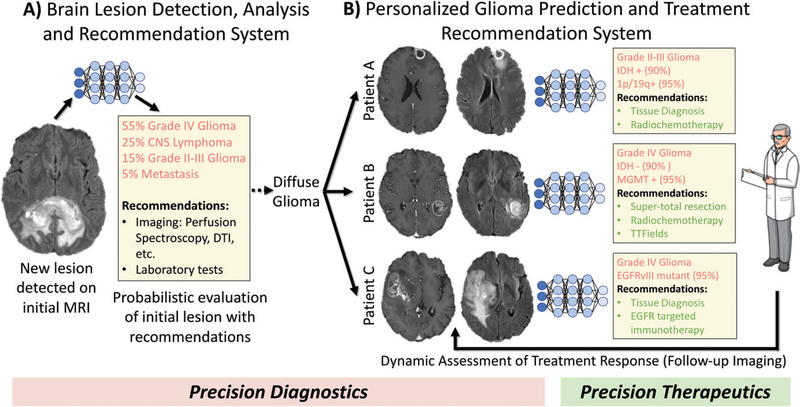
Figure 7:
Schematic of future artificial intelligence–based neuro-oncologic imaging and clinical management workflow. A, Initial lesion detection and analysis system would generate a probabilistic differential of lesion(s) seen on patient’s initial brain MR image (precision diagnostics). It would also recommend additional useful imaging examinations, laboratory tests, or tissue sampling. B, Glioma-specific module could make personalized predictions of molecular markers, survival, and treatment responses (precision diagnostics), thereby recommending optimal treatment plan(s), which would be continuously updated on the basis of follow-up imaging (precision therapeutics). CNS = central nervous system, DTI = diffusion tensor imaging, EGFR = epidermal growth factor receptor, EGFRvIII = epidermal growth factor receptor variable III, IDH = isocitrate dehydrogenase, MGMT = O6-methylguanine-DNA-methyltransferase, TTFields = tumor-treating fields.

