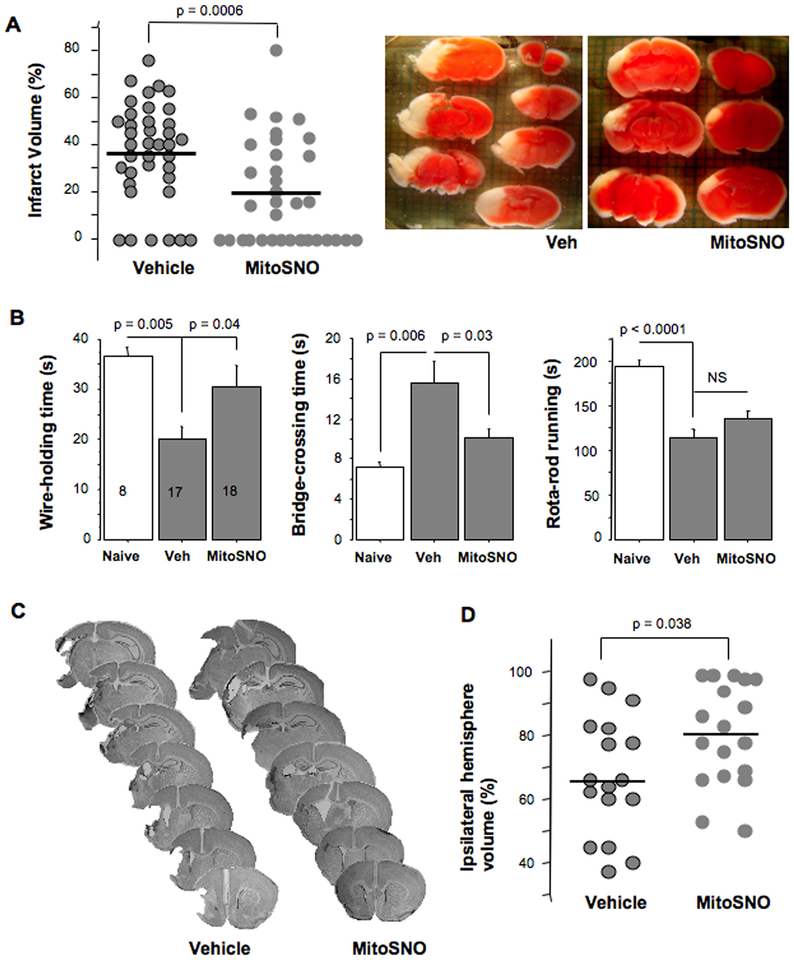
Fig. 3.
A – Actual and mean values of cerebral infarct volumes at 24 h of reperfusion and representative images of TTC-stained brains from HI-mice treated with vehicle or MitoSNO. B – Sensorimotor performance tested at ten days after HI-insult in mice treated with vehicle (Veh, n = 17) or MitoSNO (n = 18) compared to Naives (n = 8). C – Representative images of Nissl-stained coronal sections obtained at ten days following HI-insult and D – Actual and mean values of residual volume of the ipsilateral hemisphere (% of the contralateral hemisphere).

