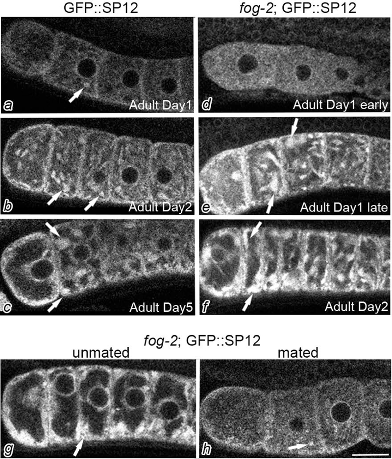
Figure 1.

Cortical ER patches accumulate in meiotically-arrested oocytes. GFP::SP12 (a-c) and fog-2; GFP::SP12 (d-f) germ lines were imaged by confocal microscopy at different developmental stages as noted and described in the Methods. The most proximal oocyte of each germ line is oriented to the left in each image of this figure and in all figures. Arrows indicate ER patches. The accumulation of ER patches in arrested oocytes is reversible as shown after mating into an unmated fog-2;GFP::SP12 female (g,h). Scale bar is 10 μm.
