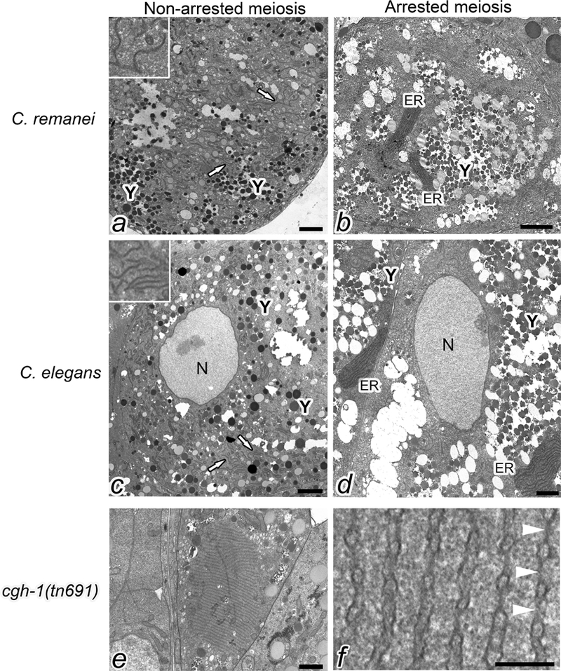
Figure 2.

ER sheets accumulate in meiotically arrested oocytes. Transmission electron micrographs of oocytes in C. remanei mated and unmated females (cross section at the cortex, lacking nuclei) (a, b), C. elegans wild type and fog-2 worms (cross section through nuclei) (c, d), and cgh-1(tn691) (cross section at the cortex) (e). The blending option in Adobe Photoshop was used to highlight the stacks of ER in b, d. White arrows in a and c indicate dispersed ER. Y indicates yolk, which occupies large areas of the cytoplasm in arrested oocytes (b, d). Scale bars are 1500 nm. The insets in a and c show higher magnification view of dispersed ER. A higher magnification view of the annulate lamellae in panel e shows nuclear pore complexes indicated by white arrowheads (f). Scale bar in panel f is 125 nm.
