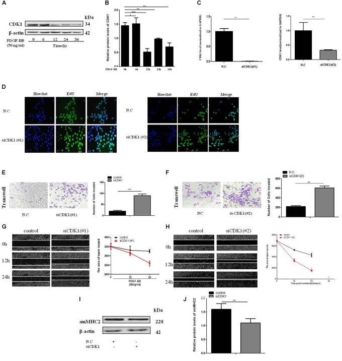
FIGURE 5.
CDK1 regulates VSMC proliferation and migration. (A) PDGF-BB (50 ng/ml) caused a time-dependent decrease in CDK1 expression levels as determined by western blot. (B) Quantitative western blot analysis of CDK1 expression in PDGF-BB-stimulated (50 ng/ml) VSMCs at various time points by western blot, data are presented as mean ± SEM. ∗p < 0.05, ∗∗p < 0.01, and ∗∗∗p < 0.001. (C) The expression level of CDK1 was detected by qRT-PCR, after transfection with siCDK1 in VSMCs, ∗p < 0.05. (D) siCDK1 increased the proliferation of VSMCs, as determined by EdU assays, scale bar = 20 μm. (E,F) Transwell were used to detect of VSMCs proliferation and migration, (Original magnification: × 200), ∗∗∗p < 0.001. (G,H) The wound closure assay was performed to investigate the migration ability of VSMCs under PDGF-BB-induced (50 ng/ml). (I) Representative western blots of VSMC phenotype marker genes transfected siCDK1 compared with control. (J) Quantitative analysis of differentiation marker gene expression in the two groups, data are presented as mean ± SEM, ∗∗∗p < 0.001.

