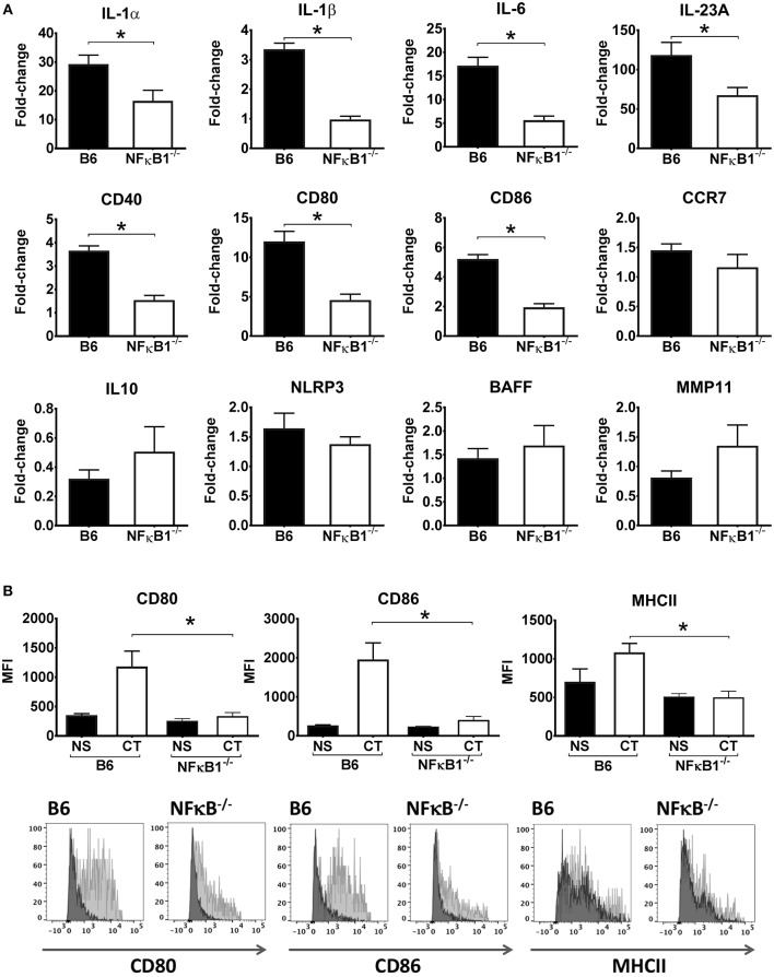
Figure 2.
Lack of NFκB abrogates CT-induced increased gene expression for pro-inflammatory cytokines and other immune activation markers in DCs. BMDCs from B6 wild-type or NFκB−/− mice were incubated in triplicates with 1 μg/ml CT for 16 h or left untreated. Purified total RNA preparations from the cells were used for inflammation focused gene expression studies by quantitative PCR. Bars represent means and SEMs of fold-change differences in gene expression between CT treated and untreated cells tested in triplicates (A). Flow cytometric analyses (B) show median fluorescence intensity (MFI) and representative FACS histogram overlays of CD80, CD86, and MHCII expression in gated BMDCs from wild-type (B6) or NFκB−/− mice incubated with either 1 μg/ml CT (light gray filled histogram) or only medium (NS), (dark gray filled histogram). *p < 0.05 for comparisons between cells treated with CT and medium alone (NS) (B).

