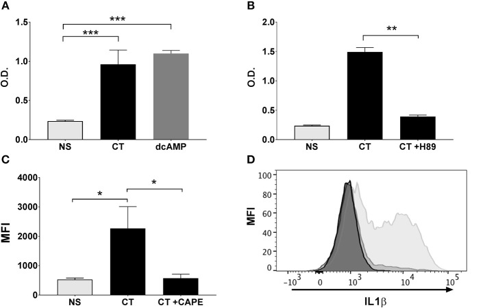
Figure 5.
NFκB activation in APCs by CT is cAMP-PKA dependent and leads to the activation of IL-1 signaling. Human monocyte cell line (THP1Blue−NFκB) were treated in triplicates with the cAMP analog dcAMP (A) or the PKA inhibitor H-89 (B) prior to treatment with 1 μg/ml CT for 16 h. O.D. is a measurement of a stable integrated NFκB inducible secreted embryonic alkaline phosphatase (SEAP) reporter construct that is directly proportional to the NFκB induction. In (C), PBMCs were treated in triplicates with or without CAPE for 1 h prior to a 16 h incubation with 1 μg/ml CT or medium only (NS), whereafter levels of intracellular IL-1β in CD14+ monocytes were analyzed by flow cytometry. Bars represent mean and SEM of median fluorescence intensity (MFI) for IL-1β. (D) shows representative ICCS histogram overlays of IL-1β expression in gated CD14+ monocytes treated with 1 μg/ml CT (light gray filled histogram), or with 1 μg/ml CT after preceding CAPE treatment (medium gray filled histogram), or with only medium (dark gray filled histogram). * represents p < 0.05 **p < 0.01, and ***p < 0.001 for indicated comparisons. Data are from one of three independent experiments showing similar results.

