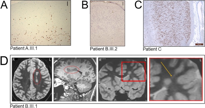
Figure 2.

Neuropathological findings. (A) In individual A.III.1, NeuN staining highlights areas of underdeveloped or focal losses of cortical layer 2 and scattered malpositioned pyramidal type neurons in cortical layer 1, consistent with FCD type Ib (original magnification 100X). Scale bar: 250 μm. (B) In individual B.III.2, Nissl‐stained section shows a laminar disorganization of the cortex indicative of FCD type Ib. Scale bar: 250 μm. (C) In individual C, NeuN staining section shows an abnormal cortical radial lamination and neurons with microcolumnar disposition consistent with FCD type Ia. No dysmorphic neurons or balloon cells were observed. Scale bar 200 μm. (D) MRI of individual B.III.1 showing deep nodular heterotopia in the left frontal lobe. Axial T2 WI (first panel from the left) and sagittal 3D T1WI (second panel from the left) show a left periventricular nodular heterotopia (in the red dashed ellipse). Coronal IR T1WI (third panel from the left) and magnification of the area included in the red square (fourth panel from the left) show a single heterotopic nodule in the deep‐periventricular left frontal white matter with the same signal intensity of the grey matter. The nodule is connected by a radial band (arrow) with the normal‐appearing overlying cortex. R = right, L = left.
