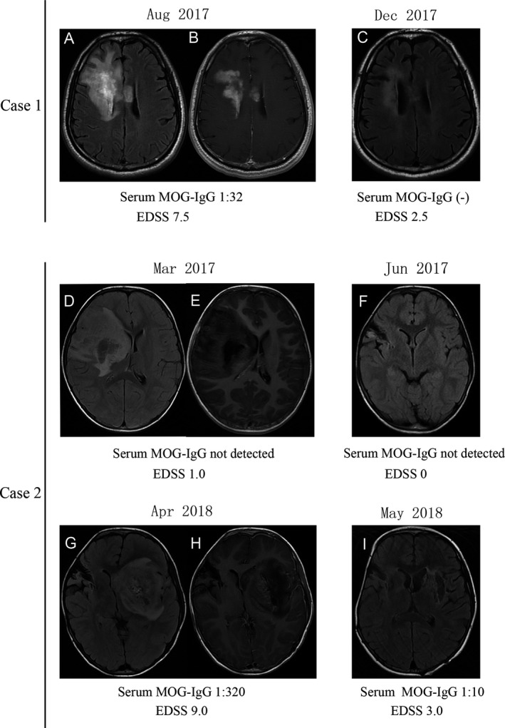
Figure 1.

(A–C) Case 1 MRI findings showed large white matter lesions with patchy Gd‐enhancement located in the right frontal lobe and basal ganglia region. (A) axial‐fluid‐attenuated inversion recovery (FLAIR), (B), Gd‐enhanced axial T1, and the lesion obviously regressed during follow‐up (C, axial‐FLAIR). (D–I) Case 2 MRI showed a large edematous lesion in the white matter of the right frontal lobe and periventricular zone with linear Gd‐enhancement (D, axial‐FLAIR, E, Gd‐enhanced axial T1) that had regressed on follow‐up at 3 months (F, axial‐FLAIR). (G–I) One year later another large edematous lesion was seen in the left basal ganglia region with patchy enhancement (G, axial‐FLAIR, H, Gd‐enhanced axial T1) that regressed during follow‐up (I, axial‐FLAIR).
