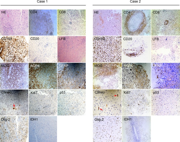
Figure 2.

In Case 1 and Case 2, both of the right frontal lobe lesions revealed extensive inflammatory cells (HE). Perivascular and parenchymal CD4+ and CD8+ T cells dominated the inflammation, with many active macrophages (CD163+). However, there was a single CD20+ B cells in the lesions of case 1 and a few CD20+ B cells in the lesions of Case 2. A marked demyelinating lesion (LFB), loss of MOG immunoreactivity, a decrease of AQP4 expression and reactive GFAP + astrocytes, and mild complement deposition (C9neo, red arrows) were also seen in the lesion. There were scattered Ki67+ cells, few cells expressed p53 or Olig‐2 proteins, and none that expressed IDH1 protein were detected in the lesions of the two cases. HE, hematoxylin and eosin; LFB, luxol fast blue; MOG, myelin oligodendrocyte glycoprotein; AQP4, aquaporin‐4; GFAP, glial fibrillary acidic protein; IDH1, isocitrate dehydrogenase 1. Magnification: HE, CD4, CD8, CD163, CD20, LFB, MOG, Ki67+, p53, Olig‐2, IDH1 × 200; AQP4, GFAP, C9neo × 400.
